
Our Research
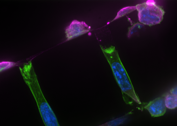
Unpicking how cancer hijacks bone
The Verbruggen Lab builds complex engineered in vitro microenvironments, such as organ-on-a-chip devices, to investigate how invading cancer cells hijack normal bone cell remodelling.
Predicting fractures in cancer patients
Using computational modelling to build digital twins from clinical scans, our team predicts the biomechanics, bone strength and fracture risk of patients who’s bones are damaged from cancer.

Seeing how bone cells sense exercise
Dr. Verbruggen’s PhD research, exploring how cells sense mechanical stimuli, in healthy and osteoporotic bone.

Understanding maternal and foetal biomechanics
Reproductive and developmental biomechanics are poorly studied areas of research. The Verbruggen Lab works with biomedical scientist and clinical collaborators to design in silico and in vitro models that help shed light on challenging research questions in these fields.
